HETA: our core platform for spatial H&E analysis

HETA (H&E Tissue Analysis) provides comprehensive tissue profiling for whole slide H&E images. It exceeds expert pathologist performance and is consistent across sites and scanners.

Validation

HETA exceeds expert pathologist performance

Validated for accuracy against an IHC-informed pathologist consensus, and for generalizability across 1,500+ images, 8 cancer types, 25+ labs and 8 scanners.

Accuracy

Matched or exceeded the performance of expert pathologists

0.74 vs 0.71

Macro F1 vs pathologist average

0.82 vs 0.78

Macro F1 on high-confidence calls

Generalizability

Consistent across primary and metastatic sites.

0.85–0.95

F1 across 8 cancer types and 5 metastatic sites

≥0.96

F1 detecting cancer cells at every site

Features

Broad coverage

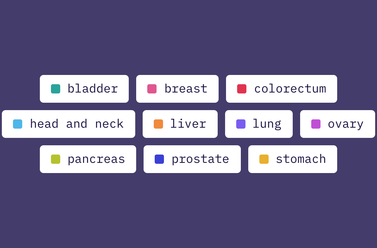
HETA currently supports analyses for ten organs.

Performance has been validated on at least 90% of invasive morphological subtypes per cancer, as well as on different tissue (e.g., primary vs. metastatic) and sample types (e.g., resections, biopsies, FNAs).

Slide, tissue, and cell analysis

Every slide passes through:

Quality control flags areas that should be excluded from downstream analyses

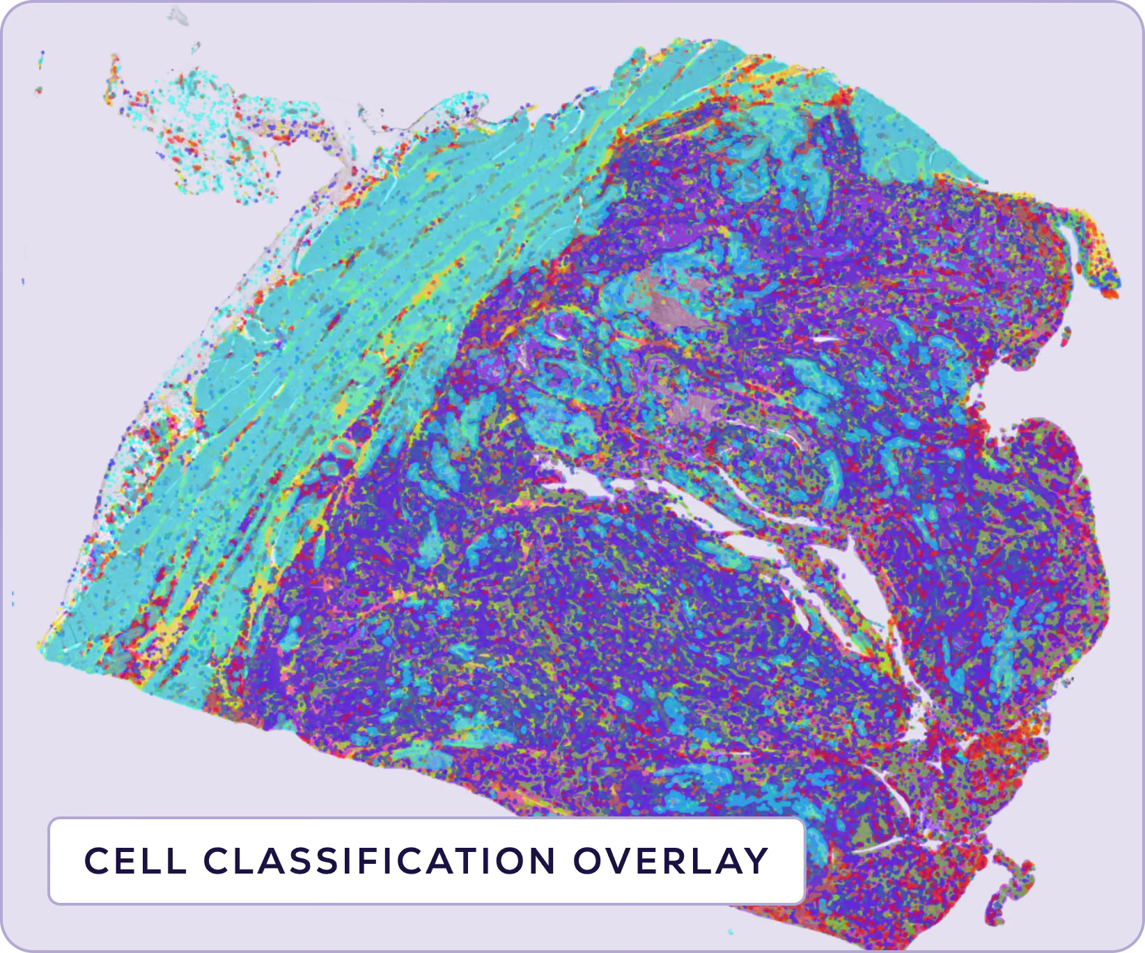
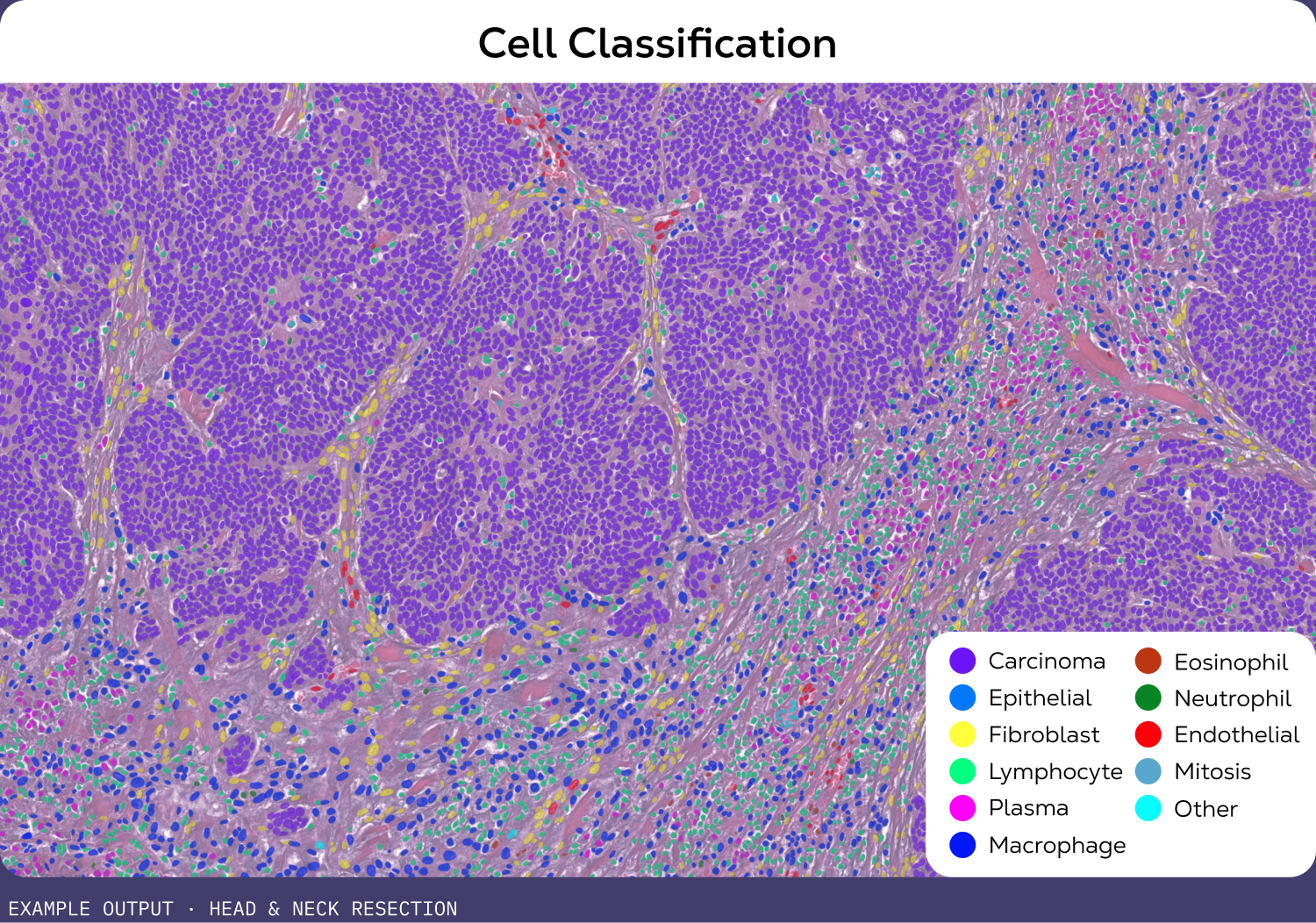
Cell detection and classification flags individual cells and classifies each into one of 11 classes

Tissue segmentation groups tissue into one of 9 classes

Confidence scores show how certain the model is about each cell classification

Tumor architecture

Map the tumor core and invasive margin, then measure what pathologists grade by eye: stroma, necrosis, desmoplasia, nuclear atypia, mitoses.

Tumor zones: core, inner and outer invasive margin, extratumoral tissue

Tumor-stroma ratio

Necrosis coverage

Desmoplasia

Nuclear morphometry

Mitotic activity and mitotic index

Immune contexture

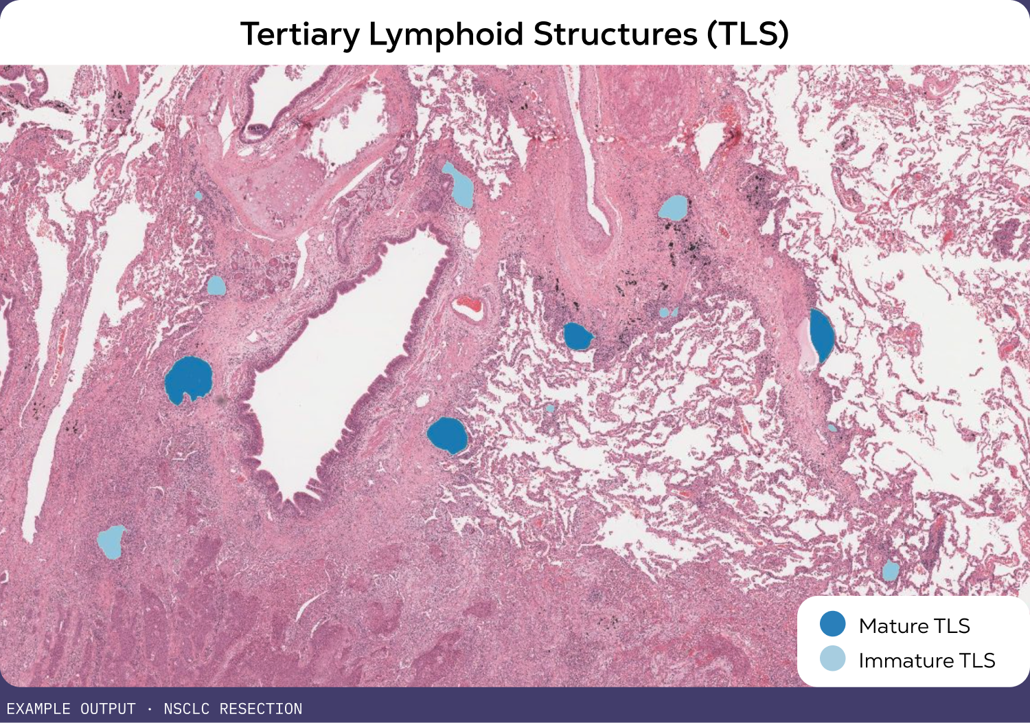
Classify immune phenotypes, count TILs and mature TLS, and score lymphocyte density where it matters, in the core and at the margin.

Tumor immune phenotypes: desert, excluded, inflamed

Tumor-infiltrating lymphocytes (TILs)

Immunoscore proxy (tumor core + invasive margin)

TLS detection and maturity classification

Mature TLS count, peritumoral vs. intratumoral

Vasculature and nerves

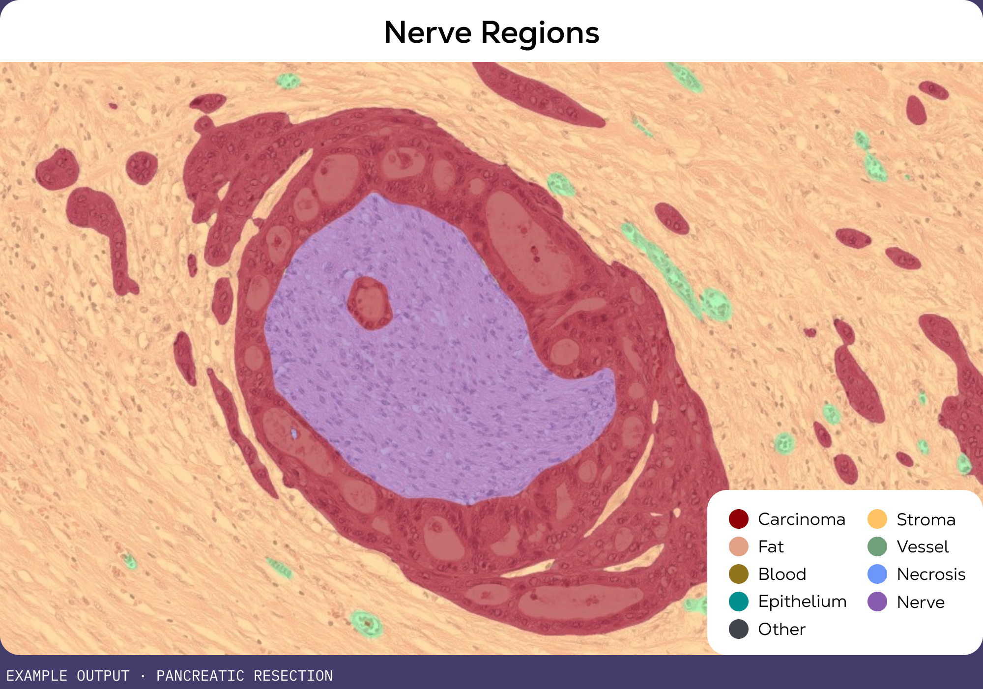
Measure vessel density and caliber, and segment nerves, for angiogenesis and invasion biology.

Microvessel density, including vessel caliber

Nerve regions

Additional readouts

Beyond the headline features, every slide comes with 20,000+ metrics at the cell, tissue, tumor zone, and neighborhood level, documented and benchmarked against TCGA.

20,000+ metrics per image

Cell, tissue, tumor zone, TLS, and neighborhood-level statistics

Readout descriptions and TCGA reference distributions

Structured for human and agent use

Documentation

HETA Product Overview

Documentation

HETA Validation Metrics

Get started with a free trial

All life sciences customers are eligible for a free trial of HETA.

Try HETA on your images, public images, or commercial images. All data will be securely transferred, analyzed, and deleted after the free trial.

HETA Inquiries
We are unable to respond to requests from personal email addresses.
Select an answer
Thank you for your submission!
Oops! Something went wrong while submitting the form.

For research use only. Not for use in diagnostic procedures.