Atlas H&E-TME Exceeds the Performance of Expert Pathologists

Today we're proud to share a new whitepaper validating Atlas H&E-TME — and we think the results speak for themselves.
H&E staining is performed on virtually every tumor specimen in pathology, but turning those slides into reliable, quantitative data at scale has remained a major challenge. Atlas H&E-TME was built to solve that, producing 4,500+ readouts per slide across eight cancer types. Now we've put it to the test.
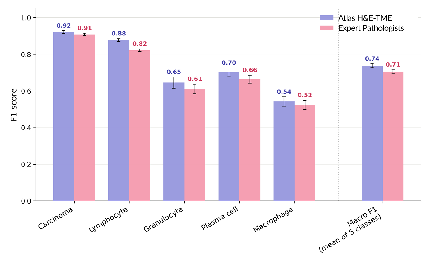
First, accuracy. H&E annotation has a known limitation: for morphologically ambiguous cell types like macrophages and plasma cells, even expert pathologists disagree. So on a small, focused cohort, we built a higher-quality reference from co-registered IHC and H&E images, with five pathologists reviewing each cell against the molecular markers to reach a consensus far more reliable than H&E alone. Measured against this standard, Atlas H&E-TME matched or exceeded pathologists working from H&E alone on every evaluated cell type.
Then, generalizability. A strong result on one focused cohort isn't enough — real-world clinical application spans many cancer types, laboratories, and scanners. So we tested Atlas H&E-TME on a separate cohort of 1,500+ cases and 200,000+ high-confidence annotations, spanning 8 cancer types, 25+ labs, and 8+ scanner models. Performance was consistently high throughout.
This is only the beginning. Through initiatives like OpenTME and continued development, Atlas H&E-TME will be expanded across more cancer types and features throughout 2026.
The whitepaper lays out the full analysis, including per-cancer-type breakdowns. It is, to our knowledge, the most thoroughly validated H&E tissue profiling system to date: built on IHC-informed, multi-pathologist consensus reference data and tested across cancer types, scanners, and labs.
Read the whitepaper here.