Atlas H&E-TME
Comprehensive analysis of the tumor microenvironment (TME)
Overview
Built using the foundation model we co-developed with Mayo Clinic, Atlas H&E-TME enables deep, accurate, and scalable spatial analyses of the TME in H&E-stained whole-slide images at single-cell resolution, empowering researchers with crucial insights for oncology drug development. Atlas H&E-TME provides qualitative polygon outputs that can be integrated into your preferred IMS, as well as thousands of higher order quantitative outputs per image.
Read the whitepaper: Atlas H&E-TME matches or exceeds expert human pathologist performance
Access Atlas H&E-TME
Atlas H&E-TME is available for both life science companies and academic researchers.
For Life Sciences
Accelerate your translational and clinical research programs with thousands of accurate, spatial features for every H&E image.
Inquire about free trialFor Academics
Access fully analyzed TCGA whole-slide images through OpenTME, or apply to run your own slides at no cost through our Research Access Program.
Explore access optionsKey advantages

Built with Atlas
Atlas H&E-TME is powered by Atlas, the industry-leading histopathology foundation model co-developed by Aignostics and Mayo Clinic.
Robust and rapid insights for every image
Atlas H&E-TME delivers comprehensive, validated analyses for cells, tissues, and higher order spatial networks, all within hours.
You control your data
Atlas H&E-TME is GDPR-compliant and developed under ISO 27001 information security standards. Your data and result sare protected during processing and transit, then automatically deleted from our network.
Features
Robust qualitative and quantitative outputs for a range of common cancer types, all within hours.
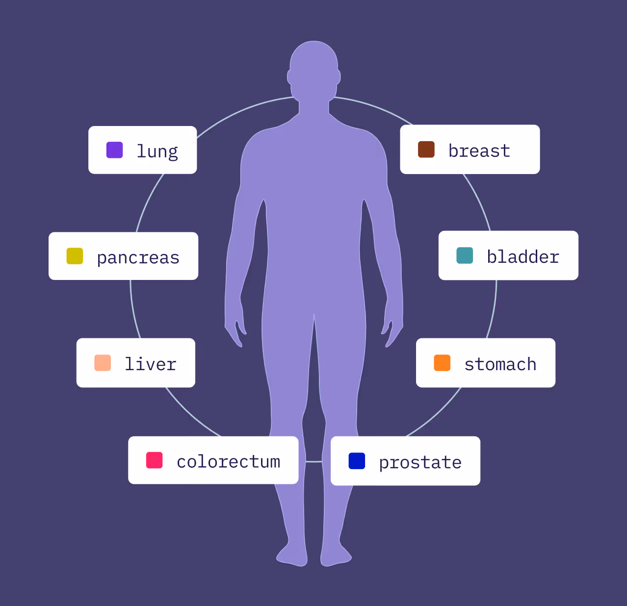
Broad coverage for common cancers
Atlas H&E-TME currently supports analyses for eight primary organs. We will continue to roll out new cancer types on a quarterly basis.
Performance has been validated on at least 90% of invasive morphological subtypes per cancer type, as well as on different tissue types (e.g., primary vs.metastatic) and sample types (e.g., resections, biopsies, FNAs).

Robust slide quality control
QC models precisely flag areas that should be excluded from downstream analyses.
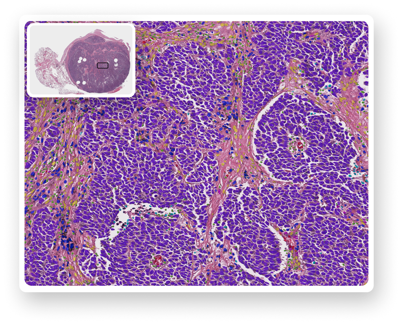
Cell detection and classification
Cell detection models flag individual cells and classify each into one of 9 different classes.
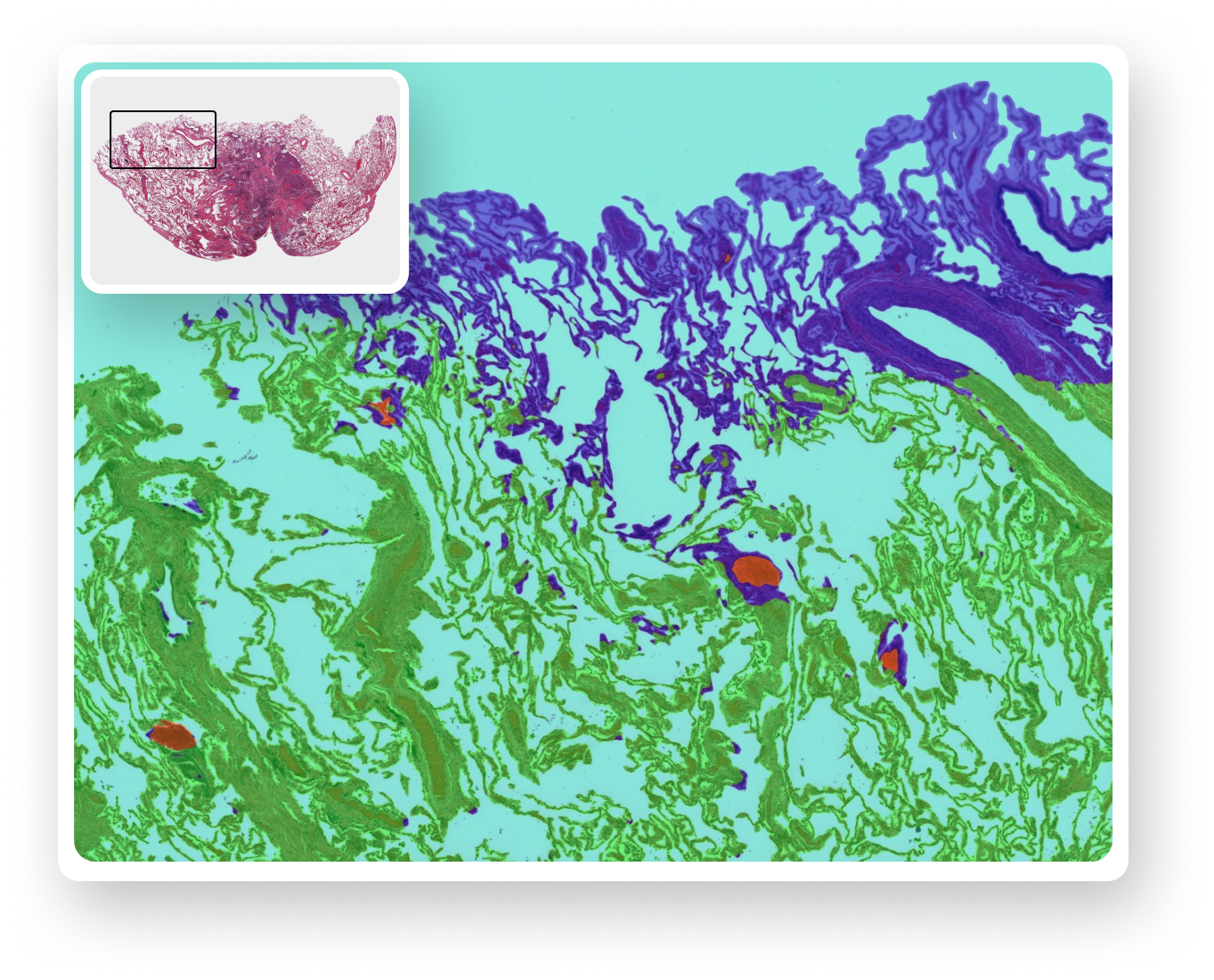
Tissue segmentation
Tissue segmentation models group relevant sections of each whole slide image into one of 7 different tissue regions.

Quantitative readouts
In addition to qualitative outputs, we provide over 4,500 quantitative metrics per image, including QC features and cell, tissue, and neighborhood-level details.

Documentation
Atlas H&E-TME Product Overview
Download our Atlas H&E-TME product summary for a detailed look at product capabilities, example outputs, and illustrative use cases of how Atlas H&E-TME is currently being used by our biopharma partners.
DownloadAtlas H&E-TME Coverage and Validation
Download our validation benchmarks summary for a detailed breakdown of Atlas H&E-TME performance data for specific cancer types, tissue segments, and cell classes. This document will be regularly updated as new indications are launched.
DownloadOpenTME Overview
Download our OpenTME overview for a detailed look at what is included in the dataset, cohort coverage, and why life sciences teams are using OpenTME.
DownloadAtlas H&E-TME is intended for research use only. Not for use in diagnostic procedures.